Academician Zhong Nanshan leads a team to publish a cover paper on STTT revealing the new mechanism of host inflammation triggered by SARS-CoV-2
2022-08-251607Recently, the team led by the academician Zhong Nanshan of Guangzhou Medical University and the team led by Professor Zhou Wenliang of Sun Yat-sen University have made an important headway in the research on the mechanism of host inflammation triggered by SARS-CoV2 (severe acute respiratory syndrome coronavirus 2). Their relevant research findings were published as an original research paper with the title of “SARS-CoV-2 nucleocapsid protein triggers hyperinflammation via protein-protein interaction-mediated intracellular Cl− accumulation in respiratory epithelium” in the international authoritative academic journal Signal Transduction and Targeted Therapy (IF = 38.104, a top journal in the Q1 of CAS) of the Nature Publishing Group (NPG). The research paper was highlighted as the cover story of that very issue of the said journal. With the aid of various technological means (including electrophysiological evaluation), the researchers found that SARS-CoV-2 nucleocapsid protein down-regulate the expression of cystic fibrosis transmembrane conductance regulator (CFTR) of the Cl- channel at the respiratory epithelium through the mechanism of protein-protein interaction and trigger intracellular Cl- accumulation, which leads to the occurrence of hyperinflammation.

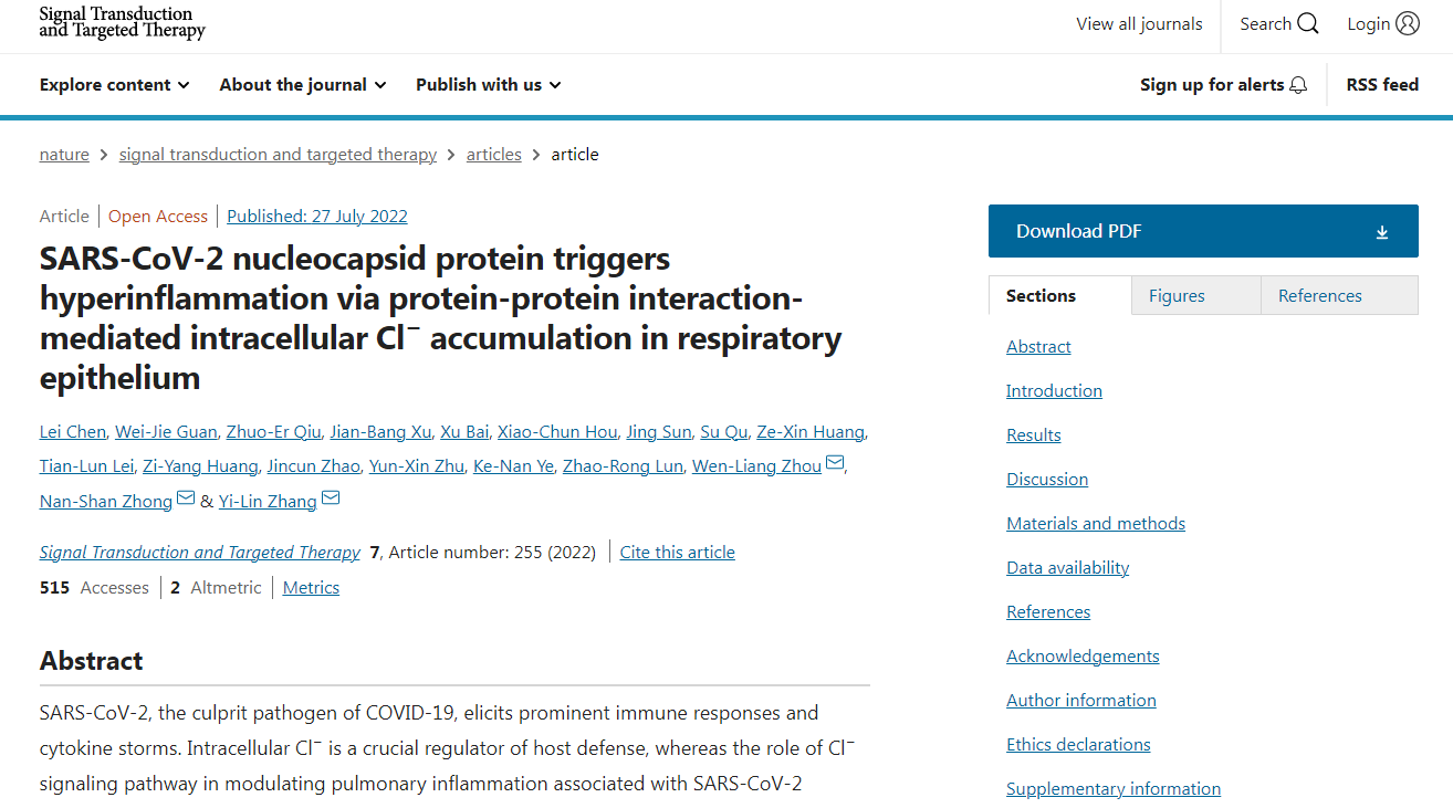
Figure 1. Screenshot of the first page of the research paper

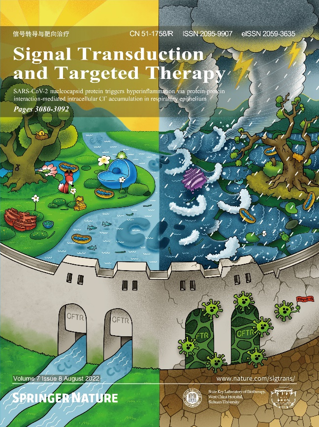
Figure 2. Cover of Signal Transduction and Targeted Therapy, Issue 8 in 2022 (in the cover picture, respiratory epithelium is depicted as a fairy land, where the river of Cl- flows through the CFTR gate on the cytomembrane wall; in the event of SARS-CoV-2 infection, the CFTR gate will be shut, resulting in Cl- accumulation and a storm of inflammatory factor)
Research background and significance
The COVID-19 (Coronavirus disease 2019) caused by SARS-CoV-2 infection has been a threat to global health. However, there is still a lack of specific drugs for COVID-19, mainly since the mechanism of respiratory damage caused by SARS-CoV-2 has not been well studied. Inflammation plays a vital role in the pathological mechanism of lung injury, which is essentially a defense reaction, a protective measure taken by the body against infection or noxious stimulation, which is of great significance in maintaining the stable equilibrium of tissues. However, if the inflammation is out of control and transforms into a continuous and excessive chronic reaction, it will cause serious adverse consequences. The current research is the first internationally reported systematic study on the mechanism of the action of intracellular Cl- signaling channel in the inflammation triggered by SARS-CoV-2 structural protein, which can provide important theoretical guidance for the exploration of new therapeutic strategies and drug development for COVID-19.
Research findings
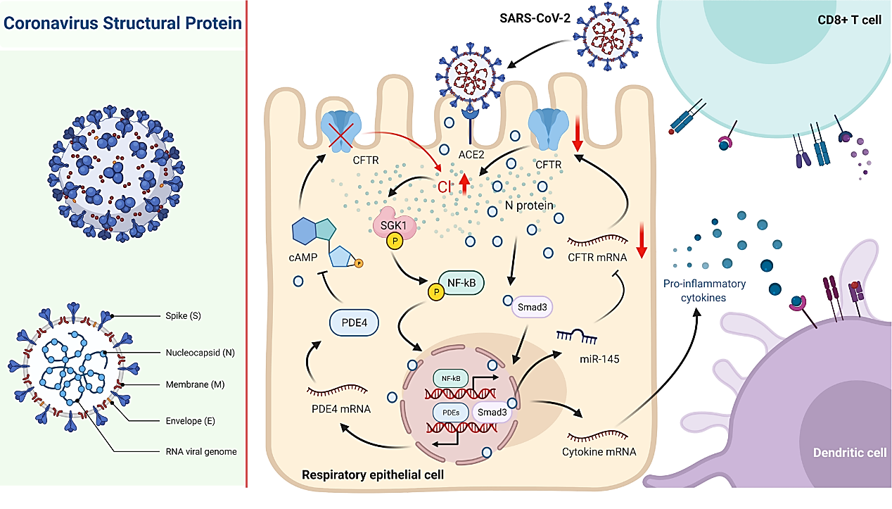
In this study, respiratory epithelium strain and primary cultured human airway epithelium were used as in vitro model, combined with the in vivo model of mice infused with viral protein in the airway and directly attacked with SARS-CoV-2, experimental technologies such as electrophysiology, molecular biology, and cell biology were applied to find that SARS-CoV-2 nucleocapsid protein stimulation can significantly inhibit the secretion of Cl- by respiratory epithelium through down-regulating the Cl- channel of CFTR. Further studies revealed that the SARS-CoV-2 nucleocapsid protein could directly interact with the Smad3 protein and mediate the down-regulation of CFTR expression through the recruitment of miRNA-145. Given that blocked transcellular Cl- secretion will probably lead to intracellular Cl- homeostasis disequilibrium, this study further found that SARS-CoV-2 nucleocapsid protein stimulation can cause abnormal increase of Cl- concentration in respiratory epithelium, which then mediated the activation of NF-κB signaling channel by activating the downstream protein sensitive to Cl- , namely serum glucocorticoid regulated kinase 1 (SGK1), thus inducing the occurrence of inflammation through micro-ion imaging technology. In addition, SARS-CoV-2 nucleocapsid protein can also reduce intracellular cAMP level by mediating the up-regulated expression of phosphodiesterase 4 (PDE4), cause the CFTR to be shut, and maintain the intracellular Cl- concentration at a high level, thus resulting in a protracted course of inflammation. On the contrary, through inhibition / knockdown of SGK1, or intervention with PDE4 inhibitor, it can inhibit the inflammation triggered by SARS-CoV-2 nucleocapsid protein stimulation.

Figure 3. The mechanism of respiratory epithelium inflammation caused by SARS-CoV-2 regulating the intracellular Cl-
Research innovation and clinical significance
In recent years, the intracellular Cl- homeostasis disequilibrium has been observed in the pathogenesis of various chronic diseases[1-6]. This study is the first attempt to systematically elaborate the mechanism of the action of intracellular chloride signaling channel in respiratory epithelium damage caused by viral infection. It is noteworthy that in 2018, in the paper published in NPG’s international authoritative journal of immunology, Mucosal Immunology, the joint research team made a groundbreaking discovery of a new type of Cl- sensing protein SGK1, and systematically interpreted the inflammation signal transduction action of intracellular Cl- in the chronic airway inflammation diseases (especially bronchiectasis) and its molecular mechanism[2]. Later, in 2019 and 2021,in another two papers published in the internationally renowned journals, International Journal for Parasitology and PLoS Neglected Tropical Diseases, the same research team further verified that the abnormal intracellular Cl- accumulation also played an important role in epithelial damage in the reproductive tract caused by parasitic infection[3-4]. Combined with the results of this study, the abnormal intracellular Cl- accumulation is common in the process of pathogen infection, which provides a solid theoretical basis for the concept proposed by the team that Cl- serves as a pathological second messenger, and the strategy to control the intracellular Cl- concentration may provide a new exploration direction for the treatment of various diseases, including COVID-19.
References
[1]Wang G, Nauseef WM. Salt, chloride, bleach, and innate host defense. J Leukoc Biol. 2015 Aug; 98(2): 163-172.
[2] Zhang YL, Chen PX, Guan WJ, Guo HM, Qiu ZE, Xu JW, Luo YL, Lan CF, Xu JB, Hao Y, Tan YX, Ye KN, Lun ZR, Zhao L, Zhu YX, Huang J, Ko WH, Zhong WD, Zhou WL, Zhong NS. Increased intracellular Cl− concentration promotes ongoing inflammation in airway epithelium. Mucosal Immunol. 2018 Jul; 11(4): 1149-1157.
[3] Xu JB, Zhang YL, Huang J, Lu SJ, Sun Q, Chen PX, Jiang P, Qiu ZE, Jiang FN, Zhu YX, Lai DH, Zhong WD, Lun ZR, Zhou WL. Increased intracellular Cl− concentration mediates Trichomonas vaginalis-induced inflammation in the human vaginal epithelium. Int J Parasitol. 2019 Aug; 49(9): 697-704.
[4] Xu JB, Lu SJ, Ke LJ, Qiu ZE, Chen L, Zhang HL, Wang XY, Wei XF, He S, Zhu YX, Lun ZR, Zhou WL, Zhang YL. Trichomonas vaginalis infection impairs anion secretion in vaginal epithelium. PLoS Negl Trop Dis. 2021 Apr 16; 15(4): e0009319.
[5] Han H, Liu C, Li M, Wang J, Liu YS, Zhou Y, Li ZC, Hu R, Li ZH, Wang RM, Guan YY, Zhang B, Wang GL. Increased intracellular Cl− concentration mediates neutrophil extracellular traps formation in atherosclerotic cardiovascular diseases. Acta Pharmacol Sin. 2022 May 5. doi: 10.1038/s41401-022-00911-9.
[6] Yang HY, Zhang C, Hu L, Liu C, Pan N, Li M, Han H, Zhou Y, Li J, Zhao LY, Liu YS, Luo BZ, Huang XQ, Lv XF, Li ZC, Li J, Li ZH, Wang RM, Wang L, Guan YY, Liu CZ, Zhang B, Wang GL. Platelet CFTR inhibition enhances arterial thrombosis via increasing intracellular Cl− concentration and activation of SGK1 signaling pathway. Acta Pharmacol Sin. 2022 Mar 3. doi: 10.1038/s41401-022-00868-9.
Original paper:https://www.nature.com/articles/s41392-022-01048-1


